ZOE Fluorescent Cell Imager
概述
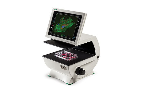
The ZOE Cell Imager simplifies fluorescence microscopy with an intuitive touch-screen interface to control the brightfield, three fluorescence channels, and integrated digital camera.
描述
The ZOE Fluorescent Cell Imager eliminates the complexities of cell imaging associated with traditional microscopes. This fluorescence imaging system combines the ease of use of a personal tablet with the power of an inverted microscope.
An Android-based platform, the ZOE Cell Imager uses an intuitive touch-screen interface to control the brightfield, three fluorescence channels, and the integrated digital camera.
The ZOE Fluorescent Cell Imager is a complete digital imaging system, allowing users to view samples, capture and store images, and create multicolor overlays. Thanks to the built-in light shield, the ZOE Cell Imager does not require a darkroom for fluorescence imaging.
Features and Benefits of the ZOE Fluorescent Cell Imager
- Simplified cell imaging — the intuitive touch-screen interface allows users to view cells, capture images, and create multichannel merges with minimal training
- Flexible operation — brightfield and three fluorescence channels enable use for routine cell culture applications and more sophisticated imaging applications
- Fluorescence imaging at your bench — light shield permits fluorescence imaging in ambient light
- Robust construction — fully integrated system with long-life LEDs, ready for intensive daily use
- LED light sources — thousands of hours of illumination that are instantly ready after power-on
- Large viewing area — the motorized stage and wide field of view allow you to see more of your sample, faster
- Small footprint — compact size accommodates crowded lab benches
Applications of the ZOE Fluorescent Cell Imager
Use the ZOE Cell Imager to check/screen samples prior to high-content analysis (HCA), high-throughput screening (HCS), confocal imaging, or fluorescence-activated cell sorting (FACS). With a brightfield and three fluorescent channels, the ZOE Cell Imager has all the features needed for daily cell culture work as well as fluorescent applications:
- Visual estimation of cell confluency
- Observation of general cell health and morphology
- Cell growth and proliferation monitoring
- Capturing cell images (with or without fluorescent labels)
- Visualization of expressed fluorescent proteins
- Immunofluorescent protein localization
- Estimation of transfection efficiency
Illumination Light Sources
- Blue channel uses a UV LED
- Green channel uses a blue LED
- Red channel uses a green LED
- Brightfield channel uses a ring of multiple green LEDs for reduced chromatic aberration
Fluorophores Compatible with the ZOE Fluorescent Cell Imaging System
This is not a comprehensive list; other dyes and fluorescent proteins with compatible excitation and emission spectra can also be used.
| Blue Channel Excitation: 355/40 nm Emission: 433/36 nm |
Green Channel Excitation: 480/17 nm Emission: 517/23 nm |
Red Channel Excitation: 556/20 nm Emission: 615/61 nm |
| PureBlu DAPI Nuclear Staining Dye |
CytoTrack Green 511/525 |
ReadiLink 555/570 Antibody Labeling Kit |
| PureBlu Hoechst 33342 Nuclear Staining Dye |
ReadiLink 492/516 Antibody Labeling Kit |
ReadiLink 594/610 Antibody Labeling Kit |
| ReadiLink 350/440 Antibody Labeling Kit |
VivaFix 498/521 Cell Viability Assay* |
VivaFix 547/573 Cell Viability Assay |
| VivaFix 353/442 Cell Viability Assay |
CFDA-SE | VivaFix 583/603 Cell Viability Assay |
| Alexa Fluor 350 | Acridine Orange | Alexa Fluor 546 |
| Alexa Fluor 405 | Alexa Fluor 488 dye* | Alexa Fluor 568 |
| Cascade Blue | BODIPY Fl* | Alexa Fluor 594 |
| CellTracker Blue | Calcein AM | Alexa Fluor 610 |
| DAPI* | DiO | Cy3* |
| Hoechst* | EGFP* | Dil Stain |
| LysoTracker Blue | ER-Tracker Green | DsRed* |
| Marina Blue | FITC* | ER-Tracker Red |
| NucBlue Fixed | MitoTracker Green FM | mCherry* |
| NucBlue Live | SYTO 9, SYTO 13, SYTO 16 | mStrawberry |
| — | SYTOX Green | MitoTracker Red* |
| — | Tubulin Green | mKate* |
| — | YFP* | RFP* |
| — | — | SYTOX Orange |
| — | — | SYTO 84, SYTO 85 |
| — | — | Texas Red* |
* R&D Tested
技术指标
| Imaging channels | Brightfield channel and 3 fluorescence channels (blue, green, and red) |
| Light source | Blue channel: UV LED Green channel: blue LED Red channel: green LED Brightfield channel: multiple green LEDs (reduces chromatic aberration) |
| User interface | 10.1 in. color (26 cm) touch-screen LCD monitor, with anti-glare and anti-fingerprint treatment, 1,280 x 768 pixel image resolution, 80–180° angle tilt range |
| Focusing mechanism | Coarse and fine, manual adjustment |
| Camera | Monochrome camera, 12 bit CMOS, 5 megapixels |
| Data format | JPEG, TIFF, or RAW image files |
| Image merge | Images from up to 4 channels can be overlaid |
| Data storage | 16 GB internal memory (~2,500 JPEG files,1,500 TIFF files, 400–800 RAW files) |
| Data export | Yes, 2 USB ports |
| Display output | Yes, 1 HDMI port |
| Objective | 20x |
| Numerical aperture | 0.40 |
| Display magnification | Standard: 175x; zoom: 700x |
| Maximum imaging area | 0.70 mm2 field of view |
| Motorized stage | 6 mm travel in X, Y direction, touch-screen control of travel speed and direction |
| Compatible with | Flasks: T25, T75, or T225 Multiwell plates: 6-, 12-, 24-, 48-, 96-, or 384-well microplates Dishes: 35 mm, 60 mm, or 100 mm Slides: chamber slides or standard glass microscopy slides |
| Software | Stand-alone Android operating system; PC is not required for operation |
| Instrument size (L x W x H) | 33 x 32 x 30 cm (13 x 12.6 x 11.6 in.) |
| Instrument weight | 9 kg (19.7 lb) |